
WHAT ARE THEY?
Mast cells are an immune cell whose function is to attack parasites in the body by releasing granules filled with histamine. They are often inadvertently triggered by pollen and environmental allergens, and can lead to allergic reaction-like symptoms (redness, itching, hives).
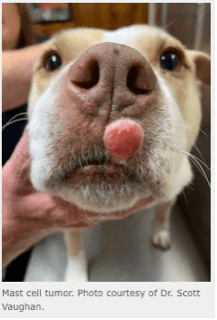
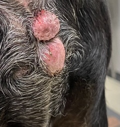
The appearance of mast cell tumors varies widely. Most are single skin or subcutaneous masses, although a small percentage of dogs have multiple lesions. Tumors composed of these cells are often reported to fluctuate in size and can become red or itchy due to the intermittent release of histamine.
Pets that have large masses or metastatic disease can also have additional signs associated with histamine release including vomiting, diarrhea, peripheral edema, fever, and rare collapse.
WHY DO THEY DEVELOP?
Mast cell tumors are the most common skin tumor of dogs. The occur mostly in older pets, and certain breeds are at increased risk including the boxer, Boston terrier, English bulldog, pug, Labrador retriever, golden retriever, cocker spaniel, schnauzer, Staffordshire terrier, beagle, Rhodesian ridgeback, Weimaraner, and Chinese shar pei.
Mast cell tumors can also occur in cats, but tend to be less aggressive compared to dogs. Regardless, these tumors are approached and treated generally in the same way as dogs.
HOW IS IT DIAGNOSED?
When a new lump is found on a dog or cat, we often perform a fine needle aspirate and cytology, where we use a small needle to obtain cells from the mass, then evaluate them under the microscope. Mast cell tumors are often able to be diagnosed immediately in-hospital, but occasionally need confirmation by sending the sample to a veterinary pathologist.
 |
 |
MY DOG WAS DIAGNOSED WITH A MAST CELL TUMOR – NOW WHAT?
“A chance to cut is a chance to cure.” There are not many cancers that we can cure with surgery alone – Mast cell tumors need to be surgically removed and sent out for biopsy. Before this happens, we like to ensure that pets are otherwise healthy to undergo anesthesia, and that they do not have evidence of metastasis (tumors that have travelled to other parts of the body). This process is called “Staging.” The level of testing performed depends on the pet’s age and health status, but usually involves bloodwork, thorough evaluation for additional skin masses, and fine needle aspirate of the nearest lymph node. Chest x-rays and abdominal ultrasound may also be recommended.
WHAT HAPPENS AFTER THE BIOPSY IS PERFORMED?
Once a mast cell tumor is removed, there are a few reasons the sample is sent to the lab for
biopsy.
- We want to see if we have “clean” margins of healthy tissue around the tumor. Mast cell tumors are notorious for spreading microscopically past the visible portion of the mass, so we like to remove at least 2cm of healthy tissue around the tumor. This is not always possible due to location or size, but is ideal.
- Grading – there are multiple methods of grading mast cell tumors, but tumors are usually divided into “low-grade” tumors, meaning they are unlikely to spread/metastasize, vs. “high-grade” tumors, which have more aggressive behavior.
MY DOG HIS MAST CELL TUMOR REMOVED AND BIOPSIED, AND IT WAS A COMPLETELY EXCISED LOW-GRADE TUMOR – NOW WHAT?
This is great news! Low-grade tumors with good histologic margins do not require any further treatment. Since there is a genetic predisposition for many dogs, routine monitoring for new masses is recommended.
MY DOG HAD AN INCOMPLETELY EXCISED OR HIGH-GRADE MAST CELL TUMOR DIAGNOSED – NOW WHAT?
There are many factors that go into formulating a plan, but surgery alone will often not prevent the spread of cancer, and we recommend consulting with a veterinary oncologist to discuss chemotherapy or radiation therapy. There are also oral chemotherapy medications that some mast cell tumors respond very well to –additional testing of the mass for “KIT mutation status” can determine if your pet is a good candidate for this medication.
CAN I PREVENT MAST CELL TUMORS IN MY DOG?
Unfortunately, most dogs are genetically predisposed, so there is no way to prevent mast cell tumors from developing. There is some weak evidence to suggest that in dogs with a history of multiple mast cell tumors, certain anti-inflammatories (NSAIDs) and control of chronic skin allergies, if present, may be beneficial.
Does your pet have a lump or a bump that is causing concern? Contact City by the Sea here to get in touch!
The links below list products proven to reduce plaque in dogs and cats:
- Veterinary Partner: https://veterinarypartner.vin.com/default.aspx?pid=19239&catId=102903&id=4952018